Designing Gamma Camera Facilities
The Hidden Infrastructure of Nuclear Medicine
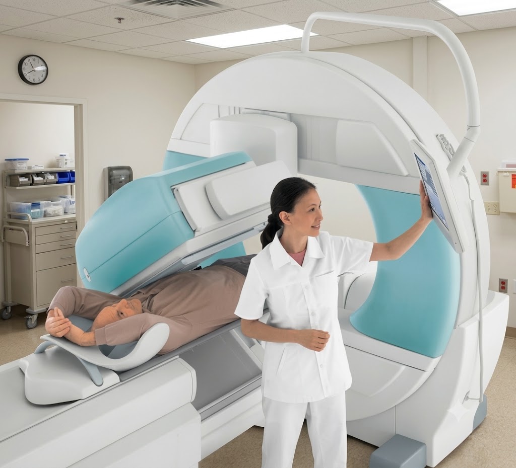
In brief
- Technetium-99m emits at roughly 140 keV, far below PET's 511 keV — but a gamma camera department images higher-energy isotopes too, and is shielded to the highest-energy study it will ever run, not the most common one.
- Iodine-131 at approximately 364 keV needs materially more shielding than technetium-99m — a structural decision that changes floor loading and can determine whether a retrofit is physically possible. The physicist gives the exact thickness; the architect must size the structure for it in advance.
- Lighter shielding removes one constraint. Every circulation and effluent constraint of a nuclear medicine department remains.
- PET-CT is a single-clock department; a gamma camera department runs multiple protocols on multiple clocks at once — same-day, multi-hour uptake, and next-day delayed imaging — so holding capacity, not camera speed, sets daily throughput.
- The multi-clock reality is mainly a capacity and scheduling problem, distinct from PET-CT's uptake period, which is largely a clinical condition of the scan.
- Most gamma cameras today are SPECT-CT hybrids, sitting under two approval paths at once — nuclear medicine provisions for the radioisotope side, diagnostic X-ray provisions for the CT component.
- Where a gamma camera joins an existing PET-CT department, the hot lab can often be shared — but only if the shared workflow doesn't break the Activity Gradient. That determination is the architect's, not the equipment schedule's.
- The regulatory review must clear before construction proceeds, and the department cannot operate without its license — the approval track runs alongside construction from day one, or it runs as a delay.
In nuclear medicine planning, the patient carries the dose, not the machine — a fact that governs both PET-CT and gamma camera departments. But a gamma camera department runs on that principle and on almost nothing else the PET-CT department relies on. The energy is different and rarely singular, the clock is longer and runs in several rhythms at once, and the planning problem that dominates shifts from shielding mass to circulation. Same family of department. Different building.
What changes when the machine is a gamma camera
The energy is lower — but not single.
Technetium-99m, the workhorse isotope of nuclear medicine, emits at roughly 140 keV — far below the 511 keV pair PET detects when a positron annihilates, and the reason a gamma camera department carries a much lighter structural shielding burden than a PET room. But a gamma camera department is rarely a single-isotope room. The same department routinely images iodine-131, gallium-67, thallium-201 and others, some of which sit well above technetium in energy.
The shielding consequence is not abstract. Iodine-131 emits at approximately 364 keV — more than twice the energy of technetium-99m — and higher energy means more shielding for the same attenuation: materially thicker barriers, which change floor loading, wall construction, and whether a retrofit into an existing structural bay is physically possible at all. The physicist’s calculation produces the exact barrier thickness for a given room geometry and activity level. The architect’s responsibility is to know that the difference exists, and to size the structure for it, before the vendor’s site-readiness document arrives — because by then the slab is poured. The shielding is therefore sized to the highest-energy isotope the department plans to handle, not the one it handles most often. A team that designs to technetium alone, because technetium is most of the workload, builds a department that cannot safely run the studies it will inevitably be asked to add. The energy question is not “how much shielding for 140 keV.” It is “which isotopes will this department ever run, and which one governs the walls.”
The clock multiplies.
PET runs on one unforgiving clock: F-18’s under-two-hour half-life, one uptake window, no slack. Technetium’s six-hour half-life is comparatively forgiving on its own — but a gamma camera department rarely runs one protocol. Different studies impose entirely different rhythms: some patients are imaged within minutes of injection, some wait two to four hours for uptake, and some studies require the patient to leave the building and return the next day for delayed imaging. The department is not a single pipeline with one clock. It is several patients on several different clocks, held in the same building at the same time, and its capacity is set by how well it holds them — not by how fast the camera itself works. This is the difference that governs the whole plan: PET-CT is a single-clock department; a gamma camera department is a multi-clock, mixed-isotope one.
The patient remains a source in more places, for longer.
An injected patient carrying activity for hours, not minutes, needs separated waiting space, toilets on the controlled side of the department with effluent still routed to decay under the same disposal rules that govern the PET-CT department, and a one-way path forward through the department rather than back through public circulation. The Activity Gradient — low-activity to high-activity, no reversal — still governs the plan exactly as it does for PET-CT. What differs is how long the patient sits inside that gradient before leaving it, and how many patients are sitting inside it simultaneously.
Why the multi-clock reality is mainly a capacity problem
PET’s uptake period is largely a clinical condition of the scan itself — the patient’s physiological state during that window affects the image. A gamma camera department’s uptake and delayed-imaging waits are mostly a different kind of problem: a capacity and circulation problem. A department holding a same-day patient, a two-hour patient, and a next-day delayed patient at once needs enough separated, comfortable holding space for all three simultaneously, or the same-day patient overflows into a two-hour patient’s seat and the whole day’s schedule compresses against itself. Hospitals that under-size this space discover it as a scheduling failure long before they discover it as a radiation-protection failure — injected patients spilling into general waiting because there was nowhere else to put them, which is among the most common operating faults in retrofitted departments in Indian private hospitals, and one no procedure can fix, because no protocol seats a patient in a chair that was never built.
One machine, two approval paths
A single machine here means two clearances, not one. The radioisotope side of the department — hot lab, injection, patient handling, waste decay — is governed by the national nuclear regulator, in India the Atomic Energy Regulatory Board, working from the Atomic Energy (Radiation Protection) Rules and the safety codes covering unsealed radioactive sources in medical use, with IAEA guidance as the international reference frame. The CT component of a SPECT-CT hybrid is a separate radiation-generating device under diagnostic X-ray provisions — a different classification, with its own room-shielding logic that has nothing to do with the nuclear medicine approval. One installation, two approval paths, coordinated and documented separately on the same drawing.
Shared hot lab: the architect’s decision, not the equipment schedule
Every gamma camera department needs a hot lab — the room where radiopharmaceutical doses arrive, are assayed and drawn, and where waste decays before disposal. Where a gamma camera is added to a department that already runs PET-CT, that hot lab and its support infrastructure can often be shared rather than duplicated — the two modalities draw on the same handling, storage and decay logic. But “shared” is an architectural decision before it is a convenience: the shared hot lab still sits at the top of the Activity Gradient, and adding a second modality’s workflow to it without re-reading the whole department’s circulation is how a working hot lab becomes a bottleneck. The vendor sizes the machine. The physicist sizes the shielding. Whether one hot lab can serve two modalities without breaking the gradient is the architect’s decision, and it is made on the plan, not in the equipment schedule. This room deserves its own full treatment, which this publication gives it separately.
What decides whether a gamma camera department works
Across projects, the decisions that separate a department that runs smoothly from one that gets corrected after occupancy are rarely about the camera itself.
- Which isotope governs the shielding — the highest-energy study the department will ever run, not the most frequent one.
- How many holding spaces, for how many simultaneous clocks — same-day, multi-hour and next-day protocols need capacity for all three at once.
- Where the hot lab sits, and whether it is shared — a shared hot lab is an economy only if the gradient survives the second modality.
- Whether next-day delayed-imaging patients re-enter through the same door as new patients — a returning, still-dosed patient crossing fresh arrivals is a gradient reversal, whatever the appointment book calls it.
- Where the injected-patient toilet’s effluent goes — the same decay principle as PET-CT, decided in section, before the slab.
The regulatory sequence does not wait for the building
The vendor’s site-readiness document describes the camera room — floor loading, access, power, environmental range. By the time it arrives, the decisions that determine the department’s fate are already frozen: where the department sits relative to public circulation, whether the injected-patient zone can be separated in the available footprint, where the effluent route falls, and whether radioactive material can arrive and decayed waste can leave without crossing a public lobby. These are concept-stage decisions. As with PET-CT, the facility design must clear the national regulator’s review before construction proceeds, and the department cannot operate without its license regardless of how complete the building is — the regulatory track runs alongside the construction track from day one, or it runs as a delay.
What this means for a hospital planning nuclear medicine
A gamma camera project looks like a smaller version of a PET-CT project and is not one. The shielding is lighter, and that removes exactly one constraint; every circulation and effluent constraint the PET-CT department carries remains, and the mixed-isotope, multi-clock reality adds its own. The vendor’s drawing describes the machine. The physicist’s calculation describes the shielding. Neither describes the department — the sequence, the separations, the shared or separate hot lab, the way an injected patient moves for hours without ever crossing the public — and that department is what the hospital is actually building. It is authored on the plan, before the machine is ordered, or the hospital ends up with a gamma camera room that cannot run its own delayed studies without breaking its hot lab’s gradient.
Studio Athenos designed the nuclear-medicine and radiation-oncology facilities at Jeevan Raksha Complete Cancer Care Centre, Bikaner — a centre on AERB’s nuclear medicine, PET-CT and radiotherapy registers, in operation.
Ar. Rahul Saxena, IGBC AP
Founding Editor · Studio Athenos, Jaipur
This article is part of Healthcare Design Dialogs, edited by Ar. Rahul Saxena, IGBC AP.