Planning a PET-CT Facility in India
How radiation safety, workflow and infrastructure become one architectural plan.
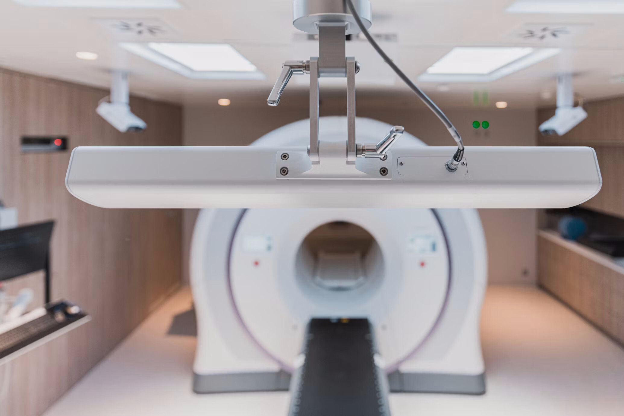
In brief
- Planning a PET-CT facility in India begins with a layout plan approved by the AERB through its eLORA system, before any construction starts — not with the scanner.
- The approved layout follows one rule, which we call the Activity Gradient: rooms are sequenced from low-activity to high-activity — reception, registration, injection, uptake, scanner, post-scan, exit — and the patient never reverses direction.
- The scanner is the least demanding part of the plan. The demanding part is F-18 FDG, the tracer, which has a half-life just under two hours and is decaying from the moment it is made — the department is the spatial translation of that clock.
- During the 45–60 minute uptake period after injection, stress and cold raise tracer uptake in muscle and fat, competing with a real lesion's signal — so the uptake room is a controlled clinical space, not overflow waiting.
- The hardest decisions are hidden in the section, not the plan: where the active toilet's effluent drain falls to a shielded decay tank, fixed before the slab is cast and effectively unfixable afterward.
- A PET-CT facility is far harder to retrofit into a running hospital than to design into a new one — the Activity Gradient, the decay-tank service path and the concrete scanner shell all need commitments an occupied building has usually already made elsewhere.
- In Tier-2 and Tier-3 India, the F-18 dose typically arrives same-day from a distant cyclotron, decaying the entire way — so the shielded receiving point and its short route to the hot lab is a site-planning decision fixed early, not a door added late.
- Studio Athenos designed the radiation-oncology and nuclear-medicine facilities at Jeevan Raksha Complete Cancer Care Centre, Bikaner, which appears on AERB's public register of licensed PET-CT centres and is in operation.
Planning a PET-CT facility in India begins with a plan approved by the Atomic Energy Regulatory Board (AERB) before any construction starts. A PET-CT department is a nuclear medicine facility that handles unsealed radioactive sources, governed by the Atomic Energy (Radiation Protection) Rules, 2004 and AERB Safety Code AERB/RF-MED/SC-2 (Rev. 2). The site and layout plan is submitted through AERB’s eLORA system and cleared for radiation safety before civil work begins, under the consenting process set out in AERB Safety Guide AERB/SG/G-3, which moves from layout approval to consent for construction to consent for operation. The approved layout arranges rooms from low-activity to high-activity: reception and general waiting at the entrance, then registration, injection, uptake rooms for injected patients, the PET-CT scanner room, a post-scan area and a controlled exit, with the hot lab and radiopharmacy at the most controlled end. A qualified Radiation Safety Officer (RSO) and a medical physicist are mandatory. A typical single-scanner PET-CT facility occupies roughly 150 to 160 square metres, with the scanner-room walls built in concrete sized to workload rather than in brick. Beyond the radiation approvals, the department must also meet NABH standards for clinical safety, infection control and patient handling. AERB clears the plan for radiation safety only; municipal and other statutory approvals are obtained separately.
What an AERB PET-CT Layout Must Show
A site and layout plan submitted through eLORA and approved by AERB before construction, with construction carried out strictly to the approved plan and any deviation reported back to AERB.
- Rooms sequenced from low-activity to high-activity, so public movement never runs toward the most active areas.
- A shielded hot lab and radiopharmacy for receiving, assaying and dispensing the radiopharmaceutical dose, positioned at the controlled end; where a Ge-68/Ga-68 generator is used, a hot lab of about 12 square metres.
- A one-directional patient route in which an injected patient does not re-enter general waiting or cross incoming patients.
- Uptake rooms for injected patients, separated from public areas as a controlled area.
- An active toilet for injected patients, with liquid effluent routed to a decay tank and held before discharge, under the Atomic Energy (Safe Disposal of Radioactive Wastes) Rules, 1987.
- A named Radiation Safety Officer and medical physicist, identified from the design stage.
- A receiving point for the delivered radiopharmaceutical dose, with a short controlled route to the hot lab.
The Planning and Licensing Sequence
- Engage a medical physicist and identify the Radiation Safety Officer at the start, not at commissioning.
- Fix the site, the dose receiving point, and the separation between controlled and non-controlled zones.
- Prepare the site and layout plan on the low-active-to-high-active principle, with one-way patient flow, hot lab, and active toilet routed to decay.
- Submit through eLORA and obtain AERB layout approval before any construction.
- Build strictly to the approved plan; report any deviation to AERB for approval.
- Move through consent for construction to consent for operation under AERB/SG/G-3, with the required radiation survey, before radioactive material is handled.
- Meet NABH requirements alongside the radiation ones.
The Scanner Is Less Demanding Than the Workflow Around It
Most promoters picture a PET-CT facility as an expensive scanner in a shielded room. The scanner is less demanding than everything around it. PET works by detecting the pair of 511 keV photons released when the tracer’s positrons annihilate in the body, a higher energy than the radionuclides used in ordinary diagnostic imaging. That is why the PET room is built in concrete rather than brick, and why AERB treats the whole department as an unsealed-source facility rather than a scan room. The CT in a PET-CT is used mainly for attenuation correction and localisation, not as a separate diagnostic room; the radiation that governs the plan comes from the patient, not the machine. For the architect, what looks like a shielding question is usually a sequencing question.
The demanding part is not the machine. It is the material. The tracer used in most PET scans, F-18 FDG, has a half-life just under two hours. From the moment it is made it is disappearing. It arrives on a clock, it decays every minute, it is injected into a patient who temporarily becomes a radioactive source, and it leaves the building as effluent that must be held until it decays. The department is the spatial translation of that clock.
The Activity Gradient
Everything in the plan follows one rule: activity rises in a single direction, and the patient never reverses it. We describe this organising principle as the Activity Gradient: the planned progression from low-activity public space to high-activity controlled space, in one direction, without reversal. AERB encodes it as arranging rooms from low-active to high-active, reception at the cold end, the hot lab at the hottest, everything between climbing in order.
Activity rises in one direction, reception to scanner, then cools toward exit. The patient enters the controlled zone at injection and is never routed back against the gradient.
Every other decision in the department is a consequence of holding that gradient: where injection sits, which corridor runs one way, where the family stops, where the drain falls. An architect who has built one reads the whole layout as this single gradient. A plan that breaks it, a dosed patient crossing a cold corridor, a waiting area placed downstream of injection, is not a detail to correct later. It is the reason a layout is returned.
What the Patient Moves Through
A patient arrives, is registered, is prepared, and is injected. At injection, the medicine changes what he is: he temporarily becomes a radioactive source and moves into the controlled zone. From this point the approved route is arranged so that he does not re-enter general waiting or cross the patients coming in behind him. He is taken to an uptake room, where he sits for roughly 45 to 60 minutes while the tracer is taken up by the tissues. Then he is scanned. Then he leaves through a route that does not run back across incoming patients, carrying residual activity that falls to negligible levels over the following hours, with discharge instructions.
That uptake period is the part no equipment plan accounts for. The patient has just been told he may have cancer, has been made radioactive, and has been separated from whoever came with him, and is asked to sit still and calm, alone, and wait. The character of that hour is set almost entirely by the plan.
Why the Uptake Room Is Not a Waiting Room
Here the patient’s condition and the quality of the scan are the same problem. During uptake, stress and cold raise uptake of the tracer in muscle and brown fat, which lights up and competes with the signal from a real lesion. A tense, cold, agitated patient tends to produce a noisier image and less stable measurements; a warm, quiet, settled patient tends to produce a cleaner one. So the calm of the uptake room is not comfort added to the medicine. It is a clinical condition of the study. An architect reads the uptake room as a controlled space, not overflow seating.
A wrongly placed uptake room is not merely inconvenient. It multiplies staff crossings into the hot zone, weakens radiation control, and cannot be relocated once the facility is licensed. The cost of placing it wrong is paid every working day for the life of the department.
The Decisions Hidden in the Section
The hardest parts of this building are not in the plan. They are in the section. The drain from an active toilet is not a drain; it is a route carrying radioactive liquid effluent, and it must fall to a shielded decay tank without passing above rooms where people sit. That vertical service zone becomes a controlled route, and it is fixed before the slab is cast. The concrete walls of the PET room are structure, not partitions. A shell drawn wrong is not corrected in fit-out; it is a structural rebuild. For the architect, the real decision here is not the wall thickness. It is where the drain falls, and that is settled in section before anything above it can be poured.
Why Existing Hospitals Struggle to Add a PET-CT Facility
A PET-CT facility is far harder to add to a running hospital than to design into a new one, and the reason is everything above. The Activity Gradient needs an uninterrupted one-way sequence, and an occupied building rarely offers a run of adjacent spaces in the right order without cutting through departments that cannot move. The decay tank and the effluent route need a vertical service path down to a safe low level, and in a finished building the shafts are already committed and the slabs already poured. The scanner-room shell must be concrete to workload, and an existing floor plate may not carry it, so strengthening it means working around live clinical areas. The dose receiving point needs a route that does not cross public traffic, which an existing entrance sequence seldom allows.
What is a set of clean decisions on a new plan becomes, in a retrofit, a set of compromises measured against structure, shafts and services that are already there. A retrofit is judged less on what the ideal layout would be and more on which of these the building can still accommodate, and sometimes on the honest conclusion that a particular floor cannot take one at all. That judgement, made early, saves a promoter from committing to a location the regulator and the physics will not allow.
The Family at the Threshold
There is a moment in every Indian PET-CT department that no room list anticipates. A patient does not arrive alone. He arrives with family, and after injection that family cannot stay with him, because he is now a source and they are not there to be exposed. That separation cannot run on signage or on staff correcting people across a corridor. It has to be built: a real boundary, and a real place for the family to wait where anxiety does not push them across it. Left to signage, that threshold becomes a daily radiation-control problem that no operator can fully police. The point where ordinary outpatient behaviour meets radioactive control is one of the places layouts most often fail, and it is an architectural problem, not a procedural one.
The Indian Condition
Most centres outside the metros have no cyclotron of their own. The F-18 dose is delivered the same day from a cyclotron in another city, decaying against its own clock the entire way, so a meaningful share of its activity is gone before it reaches the patient. That makes the shielded receiving point, and the short controlled route from it to the hot lab, a site-planning decision fixed early, not a door added late. Power is the second condition. An interruption does not only stop the scanner; it threatens the whole dosed chain and the patients already injected and waiting on a dose that will not wait. The facility is planned around continuous supply, because a failure inside the window wastes the dose, the slot and the patient’s day at once.
This is sharpest in Tier-2 and Tier-3 cities, where the promoter is often the doctor-founder, the cyclotron is furthest away, and the delivery chain is least forgiving. Dose logistics may pass through more than one vendor and more than one mode of transport, each adding delay against a clock that does not pause. Planning for that condition means designing the receiving and hot-lab end of the department for the worst delivery day, not the average one. It is the difference between a facility that runs on the days the road is slow and one that cancels its list.
Five Decisions That Decide Whether a PET-CT Facility Works
Across projects, the same small set of decisions separates a facility that is approved and runs smoothly from one that is corrected, delayed or rebuilt. None of them is about the scanner.
- Where injection sits on the Activity Gradient — it fixes the one-way route and every separation downstream of it.
- Where the drain falls. Decided in section, before the slab; effectively unfixable after.
- Where the family stops. A built boundary, or a radiation-control problem repeated every day.
- Where the dose is received. A site decision that governs how much activity survives the trip to the hot lab.
- How the scanner leaves. A removal route reserved on the first drawing, or a wall opened up later.
These are the decisions a promoter cannot see on a room list and an architect cannot add after the plan is approved.
Questions Promoters Rarely Ask Before Planning a PET-CT Facility
The questions that decide the project are usually not the ones asked first.
- Not “how big is the scanner room?” but “does the plan hold the Activity Gradient from entrance to hot lab without a single reversal?”
- Not “where is the toilet?” but “where does its effluent go, and is that route fixed in section before the slab?”
- Not “how many rooms?” but “how many patients a day, because that sets the number of uptake rooms and the rhythm of the whole plan?”
- Not “is it shielded?” but “does the layout separate cold, dosed and staff movement by plan, not by signage?”
- Not “can we start construction?” but “is the layout approved by AERB, since construction cannot begin before it is?”
The Proof
Designing and commissioning a licensed PET-CT facility exposes assumptions that drawings alone never reveal. Coordinating radiation safety, AERB approval, structure, services and clinical workflow through to an operating department is where most of the observations in this article were formed. Studio Athenos designed the radiation-oncology and nuclear-medicine facilities at Jeevan Raksha Complete Cancer Care Centre in Bikaner, which appears on AERB’s public register of licensed PET-CT centres and is in operation. A nuclear-medicine facility either passes at submission and runs, or it does not, and the difference is settled on the drawing board, before the first wall.
Infrastructure Is the Clinical Decision
Infrastructure decisions made before regulatory submission set most of the operational constraints the department will live with for the next twenty years. Designing a PET-CT facility means coordinating radiation safety, clinical workflow, structure, MEP, regulatory approval and future expansion before construction begins. That is why PET-CT planning is an architectural coordination exercise, not a room-design exercise, and why it belongs to oncology infrastructure and healthcare architecture rather than to equipment planning. The internal design of the hot lab and radiopharmacy, and the decision of whether to buy a PET-CT at all, are subjects in their own right.
For a hospital group building oncology across several sites, the PET-CT facility is the hardest unit to standardise and the most expensive to get wrong, because its constraints, the clock, the Activity Gradient, the decay of effluent, the licensing, recur at every location. Standardising it does not mean repeating one drawing; it means fixing the regulatory programme and adapting it to each site’s structure and section. Two decisions belong on the first drawing and are treated in their own right elsewhere: that the scanner must be able to leave the shell it sits in, and that the facility must be complete to regulatory standard before the first radioactive dose enters it. Both follow from the same fact this article has argued from the start.
In a PET-CT facility, the plan is not where the medicine is housed. The plan is part of the medicine.
Ar. Rahul Saxena, IGBC AP
Founding Editor · Studio Athenos, Jaipur
This article is part of Healthcare Design Dialogs, edited by Ar. Rahul Saxena, IGBC AP.